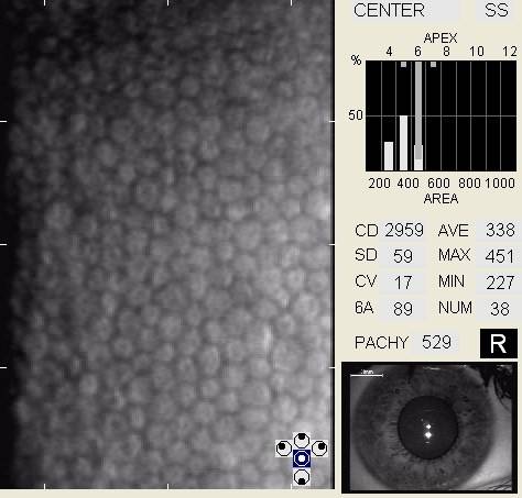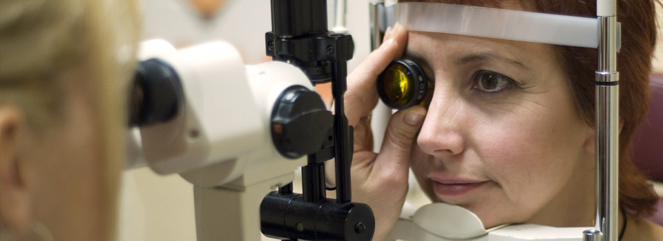
The best way to perform specular microscopy is with a modern non-contact specular microscope.
The microscope captures an image of the endothelial cells on the back surface of the cornea and the doctor analyzes the resuls to make an assessment about the health of your cornea.
Specular microscopy may be reasonable and medically necessary in the following clinical situations
- contact lens wear
- evaluation of uveitis
- evaluaton of corneal edema
- evaluation of corneal opacity
- evaluation of corneal neovascularization
- evaluation of corneal endothelial disease
- preoperative risk assessment (cataract surgery, glaucoma surgery, refractive surgery)
- postoperative surgical management (cataract surgery, glaucoma surgery, refractive surgery)


