

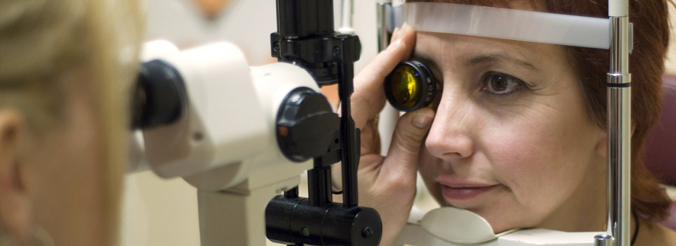
- ophthalmoscopic evaluation of the optic nerve
- fundus photographs of the optic nerve
- tonometry to measure the intraocular pressure
- corneal thickness measurements
- visual field examination
- retinal laser scans
- pupillary examination
- electrodiagnostic examination
- gonioscopy, anterior segment imaging, or ophthalmic ultrasound to evaluate the internal structures of the eye
An abnormally high IOP reading indicates a problem with the amount of fluid inside the eye. Either the eye is producing too much fluid, or it’s not draining properly.
Another method for detecting or monitoring glaucoma is the use of instruments to create images of the eye’s optic nerve and then repeating this imaging over time to see if changes to the optic nerve are taking place, which might indicate progressive glaucoma damage. Instruments used for this purpose include scanning laser polarimetry (SLP), optical coherence tomography (OCT), and confocal scanning laser ophthalmoscopy.
Visual field testing is another way to monitor whether blind spots are developing in your field of vision from glaucoma damage to the optic nerve. Visual field testing involves staring straight ahead into an instrument and pushing a button when you see a blinking light in your visual field. The test may be repeated at regular intervals so your eye doctor can determine if there is progressive vision loss.
Anterior segment imaging or ultrasound biomicroscopy may be used to evaluate how well fluids flow through the eye’s internal structures. Gonioscopy is the use of special lenses that allow your eye doctor to visually inspect internal eye structures that control fluid drainage.